Table of Contents
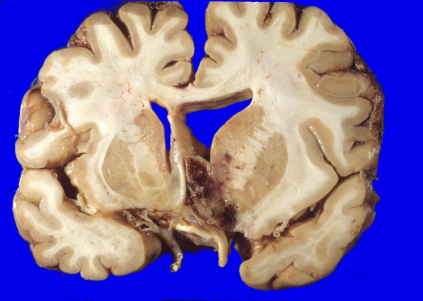
Washington University Experience | NEOPLASMS (GLIAL) | Glioblastoma - Gross Pathology | 30A1 Glioblastoma (Case 30) 4
Coronal sections through the cerebral hemispheres show a large neoplasm involving the walls of the third ventricle. The neoplasm extends from the anterior aspect of the caudate nucleus, at that point chiefly on the left, to the thalamus and basal ganglia, at this site mostly bilateral, with essential obliteration of the 3rd ventricle extending laterally to the level of the putamen and globus pallidus and posteriorly to the level of the splenium of the corpus callosum. In addition, there is a single deposit involving the roof of the 4th ventricle growing into the vermis of the cerebellum. The latter lesion is has a circumscribed appearance and appears to be in extension or a seedling and not the primary source of the neoplasm. Cross sections of the brainstem show greyish discoloration of the walls of the cerebral aqueduct, and metastatic involvement of the 4th ventricle. The ventricular system shows a slight to moderate dilation, chiefly of the left lateral ventricles.

