Table of Contents
Washington University Experience | NEOPLASMS (GLIAL) | Glioblastoma - Gross Pathology | 3A2 GBM IDHWT (Case 3) _3
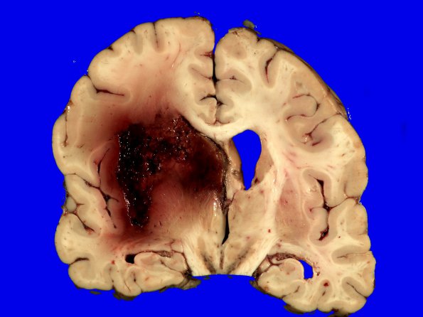
Much of the white matter and deep gray structures of the left hemisphere were taken up by a 7cm yellow-brown, hemorrhagic largely necrotic mass. More medially, there was a 4 x 4 x 4 cm acute hemorrhage centered in the white matter of the left basal ganglia and frontal lobe. There was 1.5 cm of midline shift that compressed the left ventricle completely and caused subfalcine herniation.
Not shown:
Microscopic sections of the tumor showed a high grade pleomorphic astrocytic neoplasm with extensive pseudopalisading necrosis and microvascular proliferation. There are hemorrhage and hemosiderin containing macrophages.

