Table of Contents
Washington University Experience | NEOPLASMS (GLIAL) | Glioblastoma - Gross Pathology | 5A6 Glioblastoma (Case 5) 3
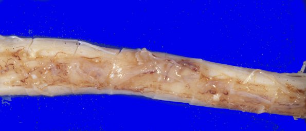
The tumor extends over the spinal cord. ---- Not Shown: Microscopic examination of the tumor showed a left temporal lobe GBM characterized by pleomorphic astrocytic cells, tumor giant cell formation, areas of necrosis, and vascular endothelial proliferation. Striking subependyrnal growth of the tumor is present throughout the ventricular system, correlating with the periventricular images which were seen on CT scan. Widespread extension into the subarachnoid space is also found, with prominent involvement of the brainstem and spinal cord meninges . Secondary invasion of cord parenchyma has apparently occurred from these meningeal foci.

