Table of Contents
Washington University Experience | NEOPLASMS (GLIAL) | Glioblastoma - Gross Pathology | 7A2 GBM (Case 7) Infiltration 2
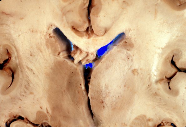
There is generalized expansion of the white matter in the left frontal lobe and in the right parietal lobe. In addition, there is an ill-defined white discoloration which appears to expand the right thalamus and measures up to 2 cm in its greatest dimension. The margins of this area blend imperceptibly with the surrounding parenchyma. ---- Serial cross sections of the brainstem and cerebellum at 3-4 mm intervals reveals recent hemorrhage in the basis pons (Duret). ---- Not shown: Microscopic sections demonstrate infiltration of all sections by a malignant astrocytic neoplasm showing extensive tumor necrosis and hence the diagnosis made was glioblastoma multiforme. The presence of tumor necrosis in the pre-radiation therapy biopsy confirms that this is indeed spontaneous tumor necrosis, and not simply a consequence of radiation therapy.

