Table of Contents
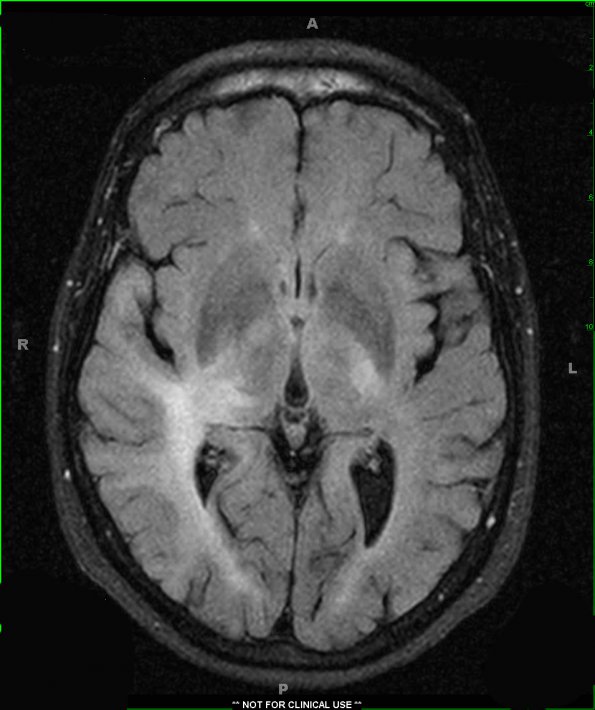
Washington University Experience | NEOPLASMS (GLIAL) | Gliomatosis cerebri | 2A1 Gliomatosis (Case 2) FLAIR - Copy
2A1,2 MRI results are shown for FLAIR (2A1) and T1 weighted image with contrast administration (2A2). FLAIR signal hyperintensities are demonstrated involving the posterior limb of the left internal capsule, pons, bilateral cerebral peduncles, and right middle cerebral peduncle. T1-weighted images showed a mildly hypointense non-enhancing process. A diffuse non-circumscribed T2 hyperintense process also expands the white matter within the right parietal, temporal, and occipital lobes.

