Table of Contents
Washington University Experience | NEOPLASMS (GLIAL) | Gliomatosis cerebri | 4A3 Gliomatosis cerebri & GBM (Case 4) MB
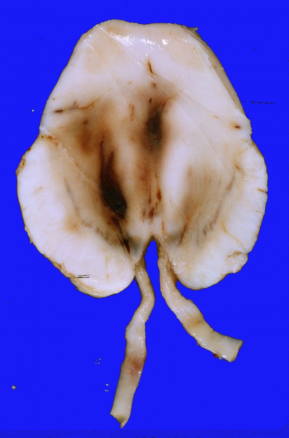
This cross section of the midbrain shows anteroposterior elongation, Duret hemorrhages within the midbrain parenchyma and also shows 3rd nerve hemorrhages on both sides, perhaps more on the right perhaps reflecting a Kernohan’s notch. Sections of the pons and medulla showed extensive hemorrhage in the tegmentum and reticular formation.

