Table of Contents
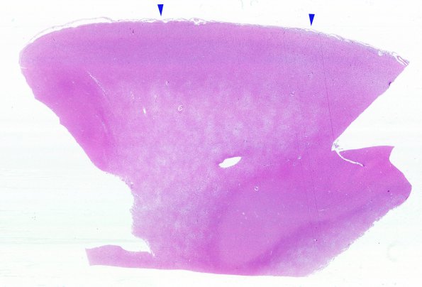
Washington University Experience | NEOPLASMS (GLIAL) | Gliomatosis cerebri | 4B1 Gliomatosis cerebri, focal GBM (Case 4) N13 WM H&E 1
4B1-3 Typical microscopic appearance of the cortical gray/white junction. Sections of the left cerebral hemisphere show a highly infiltrative astrocytic neoplasm, the majority of which is composed of low grade atypical cells with elongated and irregular hyperchromatic nuclei and scant to moderate amount of cytoplasm, percolating between the axonal fibers of white matter tracts. The pial surface is marked with arrowheads (4B1).

