Table of Contents
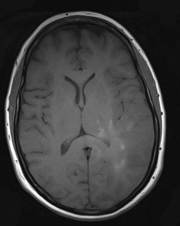
Washington University Experience | NEOPLASMS (GLIAL) | Gliomatosis cerebri | 7A2 Gliomatosis cerebri (Case 7) T1 - Copy
Multiple foci of contrast enhancement are seen within the white matter of the left temporal and parietal lobes. These areas of contrast enhancement are ringlike and primarily involving the white matter.

