Table of Contents
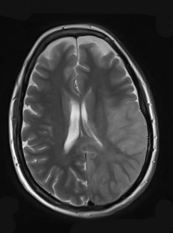
Washington University Experience | NEOPLASMS (GLIAL) | Gliomatosis cerebri | 7A3 Gliomatosis cerebri (Case 7) T2 (2) - Copy
There is increased T2 signal within the posterior aspect of the left frontal lobe, left temporal lobe, and left parietal lobe, an appearance most consistent with gliomatosis cerebri, but could also represent encephalitis, vasculitis, or nonimmune disease. This constellation of findings was thought most consistent with gliomatosis cerebri or low-grade glioma.

