Table of Contents
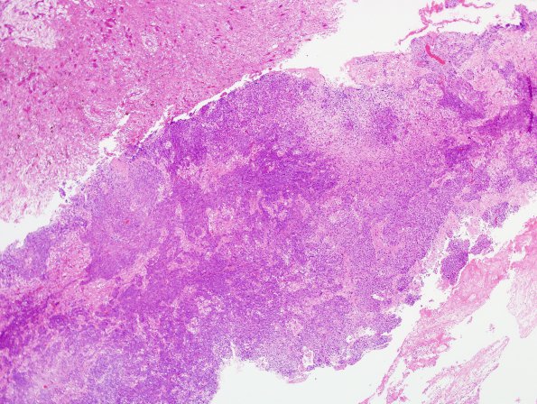
Washington University Experience | NEOPLASMS (GLIAL) | Gliosarcoma | 11A1 Gliosarcoma (Case 11) H&E 12
Case 11 History ---- The patient was a 34 year old man who underwent resection of a high grade left parietal lobe tumor in 2008 which was called a CNS PNET. FISH was performed on the first specimen and showed normal dosages (2 copies) of chromosomes 2, 7, 8, and 10. No deletions or gene amplifications were found. This genetic pattern is considered non-specific. ---- The patient was treated with a platinum based chemotherapy regimen and radiation therapy. One year later a recurrent tumor was resected. ---- 11A1,2 Sections from the recurrent resection specimen revealed multiple fragments of highly cellular, relatively solid-appearing tumor composed predominantly of small primitive cells with high N/C. There are foci of necrosis, as well as a high mitotic index and numerous pyknotic nuclei. The recurrent specimen (shown here) demonstrates foci of organizing hemorrhage and necrosis, possible related to treatment effects. However, there are also foci of residual PNET appearing tumor, some of which resemble the original specimen at least focally. Nonetheless, it was difficult to identify a definite glial or sarcomatous component by H&E histology alone.

