Table of Contents
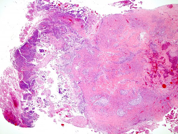
Washington University Experience | NEOPLASMS (GLIAL) | Gliosarcoma | 13A1 Gliosarcoma (Case 13) 2 H&E 2X.jpg
Case 13 History ---- The patient was an 80 year-old man presenting with memory problems and an enhancing left temporal lobe mass on MRI. The patient underwent a left craniotomy with resection of the temporal lobe tumor. ---- 13A1-4 This is a high-grade neoplasm with a biphasic tissue pattern of glial and mesenchymal differentiation. Microvascular proliferation and necrosis is present. The glial component is highly cellular and composed of pleomorphic astrocytic cells with marked nuclear atypia and increased mitotic activity. The morphology in the glial component varies greatly and includes gemistocytic areas, epithelioid areas, more primitive areas as well as smaller cells with bipolar nuclei.

