Table of Contents
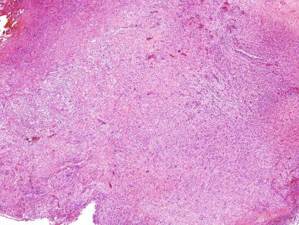
Washington University Experience | NEOPLASMS (GLIAL) | Gliosarcoma | 14A1 Gliosarcoma (Case 14) H&E 3
Case 14 History ---- The patient was a 58 year old man with a 3.5 x 1.8 cm necrotic appearing mass with enhancing margins within the left posterior parietal region and significant surrounding edema. Operative procedure: Excision. ---- 14A1-4 The neoplastic cells have a varied appearance. Much of the lesion consists of fibrillary 'bipolar' cells with thin elongate nuclei that have hyperchromatic chromatin. There are also cells with enlarged highly-irregularly shaped nuclei present. These cells have chromatin that ranges from hyperchromatic to open with prominent nucleoli. They have variable amounts of eosinophilic cytoplasm that tapers off in multiple directions. There are large swaths of necrosis with pseudo-palisading. Notice that there are delicate spindled elements separating individual large tumor cells (H&E)

