Table of Contents
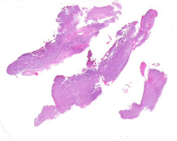
Washington University Experience | NEOPLASMS (GLIAL) | Gliosarcoma | 17A1 Gliosarcoma (Case 17) New B5 1 H&E whole mount
Case 17 History ---- The patient was a 43 year old woman who experienced headaches and seizures in mid 2007 and was found to have a right frontal lobe mass diagnosed as an oligodendroglioma, WHO Grade II with increased proliferative index. Codeletions for 1p and 19q were present. Since that time there had been interval progression with new areas of nodular enhancement by MRI. Operative procedure: Craniotomy for tumor excision. ---- 17A1-3 The neoplastic cell forms range from rare areas of oligodendroglioma with round nuclei surrounded by cytoplasmic clearing to forms with tiny bipolar nuclei and pleomorphic spindled cells. In addition there is great variability in nuclear to cytoplasmic ratios. An area of adenoid differentiation is also noted. Necrosis is evident in many portions of the tumor. The tumor cell nuclei are hyperchromatic and many have prominent nucleoli. Mitotic activity is variable throughout the tumor but in general is high. Immunostaining provided from the outside institution demonstrate that the proliferation rate, as measured by Ki-67, is very high in the tumor.

