Table of Contents
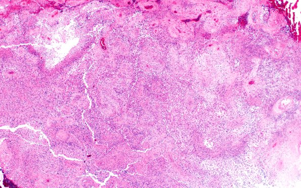
Washington University Experience | NEOPLASMS (GLIAL) | Gliosarcoma | 18A1 Gliosarcoma (Case 18) H&E 4X
Case 18 History ---- The patient was a 56 year old man with a right occipital lobe mass lesion. Clinical diagnosis: Brain tumor. Operative procedure: Excision. ---- 18A1-4 The tumor is a glioblastoma with sarcomatous features. ---- The glioblastoma is composed of small bipolar cells with atypical nuclei and a coarse chromatin pattern in a fibrillary background. Some of these neoplastic cells have abundant, eosinophilic cytoplasm. Several mitotic figures are present as well as prominent vascular proliferation and large areas of necrosis. In most areas, the vascular proliferation assumes atypical cytologic features with large nuclei, vesicular chromatin pattern, prominent nucleoli, and mitotic activity. Cells with these features form vasculocentric nodular structures that are discrete from the adjacent glial component. At one time sarcomatous areas were thought to represent progressive anaplasia of perivascular mesenchymal elements (H&E).

