Table of Contents
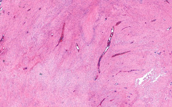
Washington University Experience | NEOPLASMS (GLIAL) | Gliosarcoma | 19A Gliosarcoma (Case 19) H&E 4X
Case 19 History ---- The patient was a 69 year old male with a left frontal lobe peripherally enhancing intra-axial mass, and an enhancing, left frontal lobe, extra-axial mass (a meningioma). ---- 19A H&E stained slides of the intra-axial mass show a high grade diffuse glioma with hypercellularity, geographic as well as pseudopalisading necrosis, and multifocal areas of endothelial hyperplasia. Individual tumor cells have moderate eosinophilic cytoplasm and oval to spindled nuclei with hyperchromasia. Tumor cells are arranged in interlacing or palisading fascicles and expand in a nodular fashion in some areas. In other areas the tumor has an infiltrative border that diffusely involves adjacent brain parenchyma, with exuberant microvascular proliferation at the border.

