Table of Contents
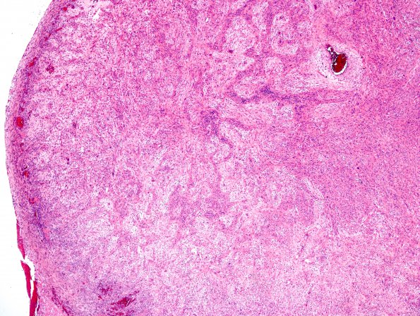
Washington University Experience | NEOPLASMS (GLIAL) | Gliosarcoma | 1A1 Gliosarcoma (Case 1) Area B 4X H&E.jpg
Case 1 History ---- The patient was a 57-year-old man who developed dizziness and headaches. MRI showed a 7.3 cm enhancing complex cystic mass in the right temporal lobe associated with uncal herniation and mass effect on the brain stem. Operative procedure: Right craniotomy with removal of temporal mass. ---- This tumor is an infiltrative malignant glioma made up of relatively small cells with moderate pleomorphism and oval to elongated nuclei with pseudopalisading necrosis, extensive geographic necrosis and microvascular proliferation. ---- 1A1-4 In many areas cells are arranged in haphazardly oriented fascicles made up of spindled cells admixed with glial elements and, as a result, the tumor takes on the appearance of a high-grade mesenchymal tumor. (H&E) ---- 1A1 This series of images represents a progressive increase in magnification in one discrete area (H&E)

