Table of Contents
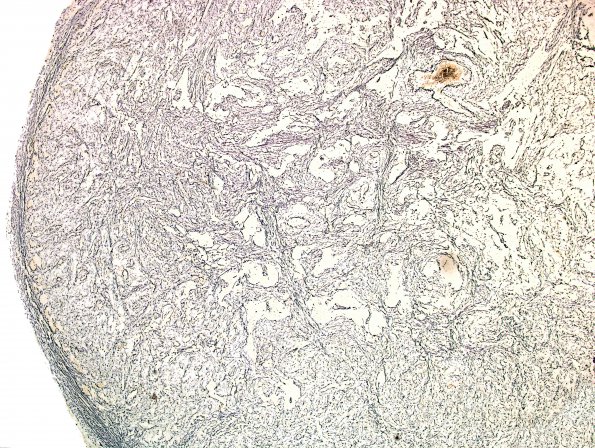
Washington University Experience | NEOPLASMS (GLIAL) | Gliosarcoma | 1C1 Gliosarcoma (Case 1) Area B 4X Retic.jpg
This reticulin stained area should be compared with the adjacent GFAP stained area shown in 1B1. The reticulin labeled areas tend to correspond to areas poor in GFAP immunoreactivity and vice-versa. Reticulin histochemistry shows high reticulin content in sarcomatoid regions.

