Table of Contents
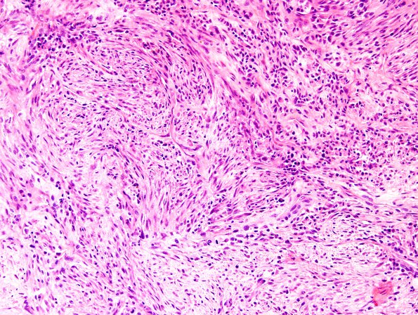
Washington University Experience | NEOPLASMS (GLIAL) | Gliosarcoma | 20A2 Gliosarcoma (Case 20) H&E 3.jpg
The specimen consists of a cellular lesion composed of variable morphologies. The predominant morphology is spindled, but areas of the tumor maintain the small, more rounded to bipolar appearance as seen in the patient's original tumor. Microvascular proliferation is identified. Geographic necrosis as well as focal areas of pseudopalisading necrosis are seen. Within the more spindled components, there is a light gray-blue myxoid-appearing background.

