Table of Contents
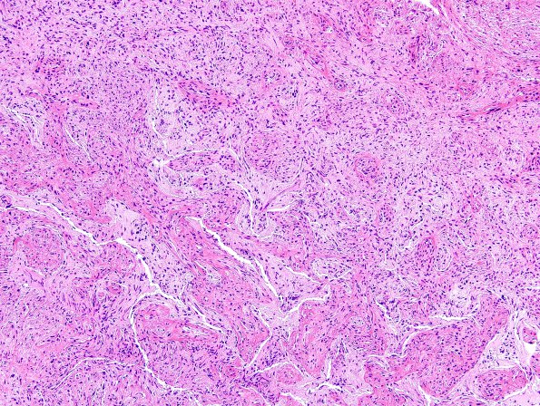
Washington University Experience | NEOPLASMS (GLIAL) | Gliosarcoma | 21A Gliosarcoma, adenoid (Case 21) H&E 14
Case 21 History ---- The patient was a 59-year-old man who presented with aphasia. MRI revealed a 6.0 x 3.2 x 5.9 cm ring-enhancing lobulated mass with surrounding vasogenic edema in the left fronto-temporo-parietal region. Operative procedure: Craniotomy. ---- 21A The specimen shows features consistent with glioblastoma including neoplastic astrocytes with bipolar or stellate morphology and hyperchromatic atypical nuclei, occasional mitotic figures, microvascular proliferation, entrapped neurons, and areas of bland (coagulative) necrosis. A second pattern, characterized by fascicles of spindled tumor cells intermixed geographically throughout most of the tumor tissue, is consistent with gliosarcoma.

