Table of Contents
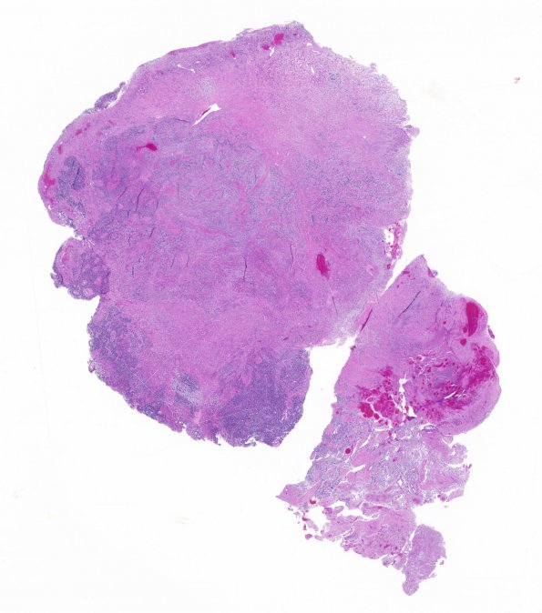
Washington University Experience | NEOPLASMS (GLIAL) | Gliosarcoma | 3A1 Gliosarcoma-PNET-Adenoid var (Case 3) H&E whole mount
Case 3 History ---- The patient was a 65 year old man who underwent surgery for a left parietal mass roughly 6 months previously, was treated with radiation/chemotherapy and underwent repeat surgery for evidence of tumor recurrence. ---- 3A1 In some areas, the tumor resembles a glioblastoma with fibrillary and gemistocytic astrocytes with a variably myxoid stromal background, mitotic activity, foci of microvascular proliferation, and necrosis. Another area resembles sarcoma, containing compact fascicles of spindled cells with moderate to marked nuclear pleomorphism and occasional bizarre multinucleated cells. Yet a third component resembles a primitive neuroectodermal tumor (PNET, see subsection in this atlas). In this section of the atlas we will focus on the gliosarcoma part. The three elements are intermingled in a fashion resembling interlocking pieces of a jigsaw puzzle. Sections from the recurrent tumor resection showed only the glial component, with numerous multinucleated giant cells. There is also extensive necrosis, calcification, and vascular hyalinization, consistent with treatment effects.

