Table of Contents
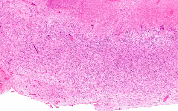
Washington University Experience | NEOPLASMS (GLIAL) | Gliosarcoma | 7A1 Gliosarcoma (Case 7) B H&E 4X
Case 7 History ---- The patient was a 71 year old woman with mental status changes. MRI showed a 2.6 cm thick walled rim enhancing mass in the left frontal lobe with intense vasogenic edema and restricted diffusion. Operative procedure: Image-guided microscope-assisted left frontal craniotomy. ---- The following images show alternating low (4X) and higher (20X) magnification images of adjacent sections of one block of tumor stained for a variety of elements. ---- 7A1,2 Sections show a highly cellular neoplasm composed of a moderately pleomorphic population of cells composed predominately of smaller bipolar cells as well as a more elongated to spindled cell component, areas of extensive vascular proliferation and necrosis. (H&E)

