Table of Contents
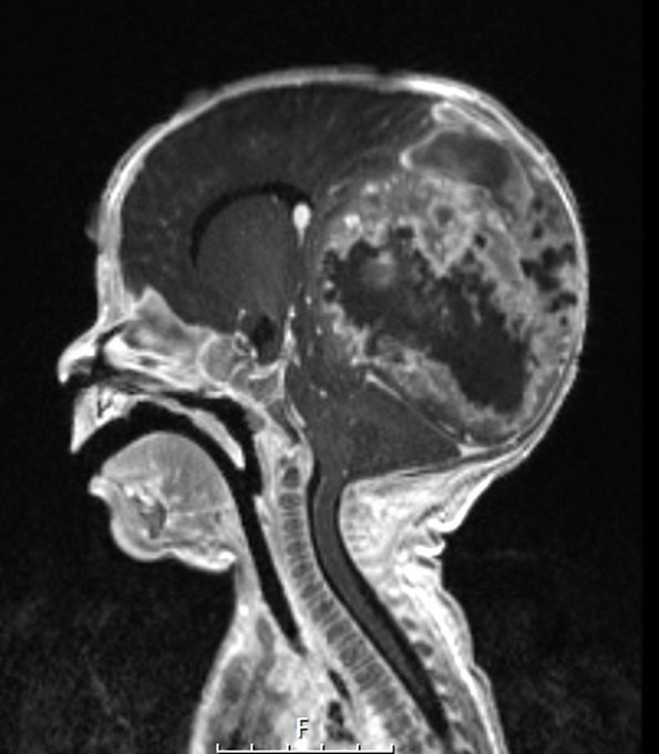
Washington University Experience | NEOPLASMS (GLIAL) | Infant-Type Hemispheric Glioma | 1A1 Infant-Type Hemispheric Glioma (Case 1) T1W sagittal - Copy
Case 1 History ---- The patient was a 7-week-old full-term male who presented with emesis and pale and shaky episodes to an outside ER. Cranial ultrasound showed a grade IV hemorrhage. He was transferred to SLCH where imaging showed a 9.3 x 8.7 x 7.2 cm heterogeneous, hemorrhagic, enhancing mass in the right parietal and occipital lobes. Operative procedure: brain biopsy. ---- 1A1,2 This T1-weighted contrast administered massive tumor is shown in sagittal (1A1) and axial (1A2) scans.

