Table of Contents
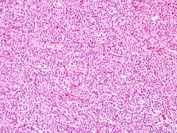
Washington University Experience | NEOPLASMS (GLIAL) | Oligodendroglioma - Histologically Defined (most del 1p19q) | 10A1 Oligodendroglioma (Case 10) H&E 7
Case 10 History ---- The patient was a 42 year old woman who presented with witnessed grand mal seizures. CT imaging showed a predominantly hypodense left frontal mass lesion with patchy hyperdensities consistent with calcifications. Operative procedure: Left frontotemporal craniotomy with excisional biopsy. ----
Sections show cortex and subcortical white matter involved by a hypercellular glial neoplasm growing in sheets. Tumor cells have round to mildly ovoid nuclei with open and finely stippled chromatin, peri-nuclear cytoplasmic clearing and fine vasculature with a chicken-wire pattern. In areas the vasculature has an increased density but remains single-walled without frank endothelial hyperplasia. Mitotic figures are sparse and numbers up to 1/10HPF. Necrosis is not present. Portions of the lesion show calcifications.

