Table of Contents
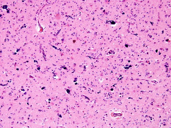
Washington University Experience | NEOPLASMS (GLIAL) | Oligodendroglioma - Histologically Defined (most del 1p19q) | 10A2 Oligodendroglioma (Case 10) H&E 5
Sections show cerebral cortex and subcortical white matter involved by a hypercellular glial neoplasm growing in sheets. The neoplastic cells have a regular distribution. They have round to mildly ovoid nuclei with open and finely stippled chromatin; many of the cells show peri-nuclear cytoplasmic clearing. Background vasculature is fine and shows a chicken-wire pattern. Mitotic figures are sparse and number up to 1/10HPF. Necrosis is not present. Portions of the lesion show calcification often outlining capillary walls. Not Shown: Immunohistochemical stains performed by the referring institution include synaptophysin which shows an infiltrative growth pattern. Ki67 demonstrates a labeling index of 5.7%. P53 shows weak scattered positivity in <1% of neoplastic cells. FISH studies show co-deletion of both chromosome 1p and 19q.

