Table of Contents
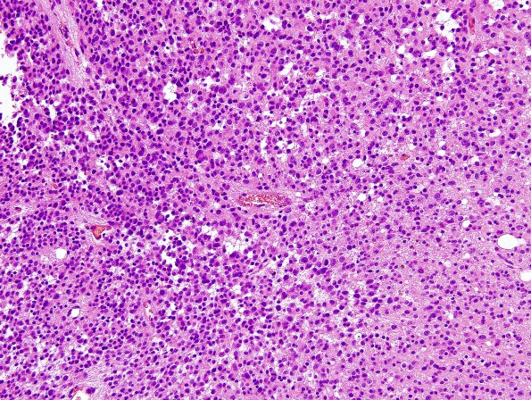
Washington University Experience | NEOPLASMS (GLIAL) | Oligodendroglioma - Histologically Defined (most del 1p19q) | 11A1 Oligodendroglioma WHO-III (Case 11) minigemistocytes H&E 1
Case 11 History ---- The patient is a 42 year old man with a previous diagnosis of oligodendroglioma, WHO grade II with codeletion of chromosomes 1p and 19q. He is status-post resection in 2003, and treatment with Temodar chemotherapy. To date he has had no radiation. MRI of the brain shows findings worrisome for recurrent right frontal oligodendroglioma at the medial surgical margin. Operative procedure: Right frontal craniotomy with tumor resection and intraoperative MRI. ---- 11A1-4 Sections show a hypercellular glial neoplasm consisting of neoplastic cells distributed with a regular, sheet-like pattern. Portions of the lesion show formation of microcysts. The neoplastic cells have relatively round nuclei with mild shape irregularities, variably open chromatin with areas of chromatin clumping, and occasional rims of peri-nuclear cytoplasmic clearing (peri-nuclear 'halos'). 'Mini-gemistocytes' with plump eosinophilic cell bodies and eccentrically placed nuclei are also present. The background vasculature is fine caliber and does not show endothelial hyperplasia. Necrosis is not identified. Multiple portions of the specimen show foci of increased cellularity, increased nuclear atypia, and increased mitotic activity. Mitotic counts show foci where the mitotic index ranges from 5 to 7 mitoses/10HPF.

