Table of Contents
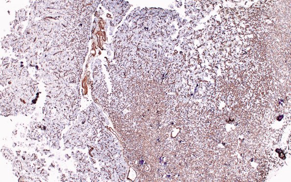
Washington University Experience | NEOPLASMS (GLIAL) | Oligodendroglioma - Histologically Defined (most del 1p19q) | 13A1 (Case 13) Oligo Coll IV 20X
Case 13 History The patient is a 23 year old man with a brain tumor. Operative procedure: Craniotomy for tumor resection. Clinical diagnosis: Left frontal lobe tumor. ---- The neoplasm is composed of hypercellular sheets of oligodendroglial cells with enlarged pleomorphic nuclei that diffusely infiltrates cortex. Many of the neoplastic cells show the "fried egg" appearance typical of oligodendroglial cells, however, in other areas the cells have a small rim of eosinophilic cytoplasm. The neoplastic cells are surrounded by a network of delicate capillaries and abundant microcalcifications are present. Also, focal microcystic change is identified. Features of mild anaplasia demonstrated by the tumor include increased mitotic activity and focal marked hypercellularity. Immunohistochemistry shows that the neoplastic cells are diffusely positive for S100 and are GFAP negative. ---- 13A1-3 Type IV collagen highlights the extensive microvascular network.

