Table of Contents
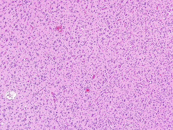
Washington University Experience | NEOPLASMS (GLIAL) | Oligodendroglioma - Histologically Defined (most del 1p19q) | 15A1 Oligodendroglioma, Grade 2 1p19q del (Case 15) H&E 3
Case 15 History ---- The patient is a 68-year-old man with a recent seizures and a left frontal T2 bright, non-enhancing mass on MRI suspicious for glioma. Operative procedure: Craniotomy. ---- 15A1-4 Sections show a moderately cellular infiltrative glial neoplasm with occasional microcalcifications. There is both cortical and white matter involvement with a prominent microcystic growth pattern. There is mild nuclear pleomorphism, with the majority of tumor cells containing round to oval nuclei with delicate chromatin, small nucleoli, and clear perinuclear halos. A mucin-rich background is seen in some areas. There is prominent formation of secondary structures with tumor satellitosis around neurons and subpial tumor aggregates. Occasional mitotic figures are seen with 2 mitoses/10HPF. There is no microvascular proliferation or necrosis.

