Table of Contents
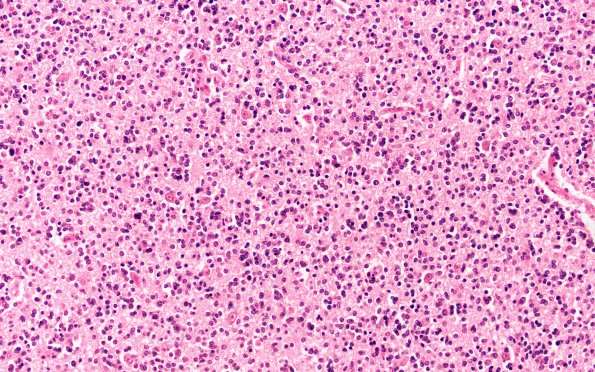
Washington University Experience | NEOPLASMS (GLIAL) | Oligodendroglioma - Histologically Defined (most del 1p19q) | 1B Oligodendroglioma (Case 1) H&E 20X
1B Microscopically, there was a diffuse subtle infiltration of the gray and white matter by tumor cells with largely round nuclei and a hint of perinuclear clearing. Mitotic figures were scarce. The tumor cells tended to cluster around neurons and in a subpial location. The infiltrate involved the left and right frontal lobes, inferomedial portions, hippocampus, unci, basal ganglia, calcarine regions, the left thalamus and internal capsule, the mesencephalon (especially the tectum), the pontine tegmentum, and extending caudally to the distal medulla. ---- Comment: This tumor was diagnosed in 1968 as an oligodendroglioma but we have been unable to find the blocks for a definitive modern immunohistochemical and FISH workup. The 9 year course of the tumor does suggest an indolent course not inconsistent with an oligodendroglioma but it remains unproven.

