Table of Contents
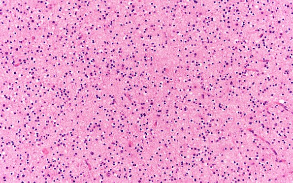
Washington University Experience | NEOPLASMS (GLIAL) | Oligodendroglioma - Histologically Defined (most del 1p19q) | 3B Oligodendroglioma (Case 3) H&E N16 20X 1
Microscopic examination of the right temporal lobe tumor mass reveals a moderately cellular tumor which was interpreted at the time as a mixed glioma with both astrocytoma and oligodendroglioma components. The astrocytic component was characterized by tumor cells with mild hyperchromatic and moderately pleomorphic oval to irregular nuclei which were associated with a small amount of eosinophilic fibrillary cytoplasm. In the oligodendroglioma component, the tumor was characterized by round to oval nuclei with delicate chromatin and inconspicuous nucleoli which are associated with scant cytoplasm. Some tumor cells have typical perinuclear halos, resulting in a "fried egg" appearance. Mitoses were difficult to find. No evidence of vascular proliferation or tumor necrosis was seen. A radiation effect is also noted including the hyalinization of blood vessel walls and focal parenchymal calcifications.

