Table of Contents
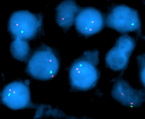
Washington University Experience | NEOPLASMS (GLIAL) | Oligodendroglioma - Histologically Defined (most del 1p19q) | 7B1 Oligodendroglioma -1p 1q (Case 7B1)-2 cropped - Copy
Case 7B1,2 History ---- The patient is a 37 year old man with a non-enhancing frontal lesion. Sections show an infiltrative glioma composed of cells with round irregular nucleoli, many with surrounding perinuclear clear zones/halos. Delicate capillaries are detected in many areas of the lesion, and there is focal microcystic change. The tumor infiltrates into the overlying cerebral gray matter with perineuronal and perivascular satellitosis and subpial accumulation of tumor cells. Mitotic figures are infrequent, and there is no evidence of endothelial hyperplasia or necrosis. Overall, these features are those of an oligodendroglioma, WHO grade 2. FISH studies show loss of 1p (7B1) and 19q (7B2).

