Table of Contents
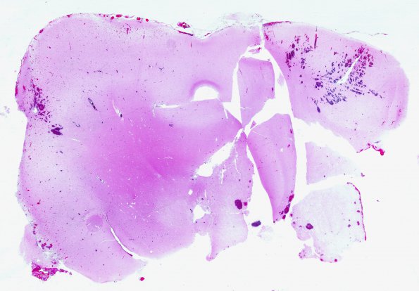
Washington University Experience | NEOPLASMS (GLIAL) | Oligodendroglioma - Histologically Defined (most del 1p19q) | 9B1 Oligodendroglioma, focal anaplasia (Case 9) WM
9B1-3 Sections of the right frontal tumor show a hypercellular, infiltrating glial neoplasm composed of cells with round to oval nuclei, crisp nuclear membranes, and a moderate amount of clear to eosinophilic cytoplasm consistent with oligodendroglioma. Some of the cells have abundant eosinophilic cytoplasm with the nucleus displaced, consistent with mini-gemistocytes. There are secondary structures consisting of perineuronal and perivascular satellitosis. Mitoses are easily identified with a count of 10mitoses /10 HPF. There are scattered microcalcifications. There is no necrosis or endothelial hyperplasia. The tumor extends into the leptomeningeal space. In this particular case, there were deletions of both 1p and 19q.The histology is consistent with that of an oligodendroglioma with focal anaplasia, Grade 3.

