Table of Contents
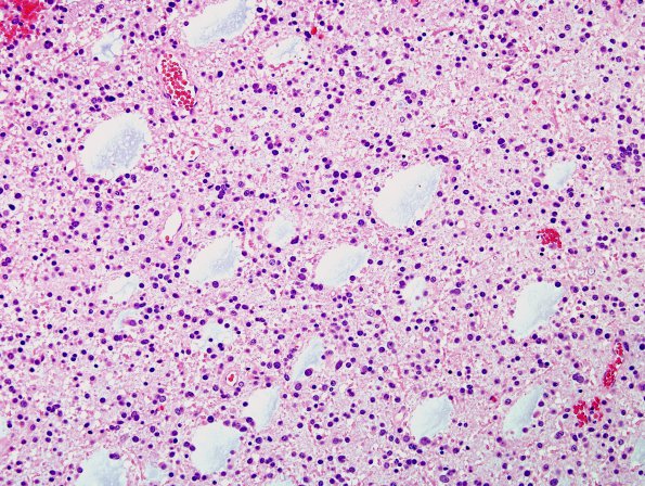
Washington University Experience | NEOPLASMS (GLIAL) | Oligodendroglioma - IDH1 mutant, del 1p19q - Grade 2 | 19 Oligodendroglioma, WHO II (Case 19) H&E 2
Case 19 History ---- The patient is a 49 year old woman who had fallen and struck her head in July 2009. A subsequent CT scan showed a left posterior frontal lesion that was confirmed on MRI which demonstrated a non-enhancing mass with increased T2 signal and FLAIR. She was managed by following due to the tumor location (eloquent cortex) until 8/2011 when she had a generalized tonic-clonic seizure. At this time the decision to operate was made. Operative procedure: Left frontal
craniotomy with IMRI, stealth guidance and motor mapping for tumor excision. ---- Sections show a cellular neoplasm composed of sheets of cells with prominent areas of microcystic degeneration. A delicate vascular network is seen within the tumor. The majority of the tumor cells have rounded nuclei and occasional perinuclear clearing. There are abundant cells with a pericentral expansion of cytoplasm representing mini-gemistocytes. Mitotic activity overall is low but focally reaches 2/10 high powered fields. ---- Not shown: Immunohistochemistry for a mutated form (R132H) of isocitrate dehydrogenase 1 (IDH1) is positive in the tumor cells. There are scattered p53 weakly positive cells but the vast majority are negative. Glial fibrillary acid protein (GFAP) largely excludes the tumor cells, however, some gliofibrillary oligodendroglial forms are present. The proliferation index overall is low reaching up to 3.7% measured by immunostaining for Ki-67.

