Table of Contents
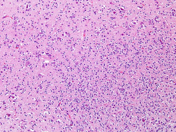
Washington University Experience | NEOPLASMS (GLIAL) | Oligodendroglioma - IDH1 mutant, del 1p19q - Grade 2 | 1A1 Oligodendroglioma (Case 1) H&E 1
Case 1 History (S11-5840) ---- The patient is a 52-year-old man with a history of new onset seizures in November of 2010. Magnetic resonance imaging shows a 5.2 x 2.2 x 2.2 cm area of increased T2/FLAIR signal and decreased T1 signal, with two subtle areas of postcontrast enhancement, located within the anterior left frontal lobe, involving several adjacent gyri. Radiological interpretation: Primary glial neoplasm such astrocytoma or oligodendroglioma. Operative procedure: Left frontal craniotomy with tumor resection. ---- 1A1-4 Histological sections show multiple pieces of neocortex and subcortical white matter involved by a diffuse glial neoplasm at variable density.

