Table of Contents
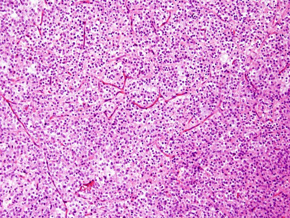
Washington University Experience | NEOPLASMS (GLIAL) | Oligodendroglioma - IDH1 mutant, del 1p19q - Grade 2 | 4A1 Oligodendroglioma (WHO-Grade 2,Case 4) H&E 1
Case 4 History ---- The patient is a 47 year-old man with a history of oligodendroglioma WHO grade 2 (1p/19q deletion). Per clinical record in 4/2009, the patient has been treated on the observation alone arm of RTOG 9802. MRI of the brain (4/2011) shows interval increase in size of a nodular T2 hyperintensity with diffuse restriction, consistent with a slow interval growth of tumor. Operative procedure: Awake craniotomy with use of IMRI for tumor excision. ---- 4A1-3 Sections show an infiltrative hypercellular glial neoplasm comprised of neoplastic cells with oligodendroglial features. The cells have round nuclei with mild contour irregularities, and chromatin with variable clearing and stippling. Many of the neoplastic cells show peri-nuclear cytoplasmic clearing and have a so-called 'fried egg' appearance. The vasculature is fine and of thin caliber, shows 120 degree angle branching, i.e. shows a so-called 'chicken wire' appearance. Microcyst formation is present. Portions of the specimen show increased cellularity and crowding, and also have cells that show increased nuclear contour irregularities and hyperchromatic chromatin. The vasculature within these areas remains single-layered. Necrosis is not identified. Mitoses are present and focally number up to 2/10HPF.

