Table of Contents
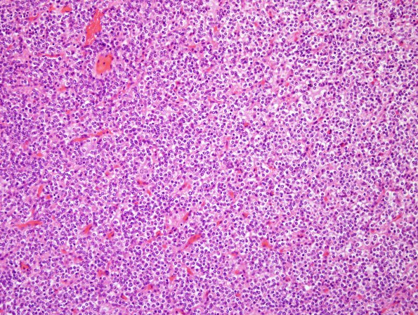
Washington University Experience | NEOPLASMS (GLIAL) | Oligodendroglioma - IDH1 mutant, del 1p19q - Grade 2 | 5A1 Oligodendroglioma, WHO 2 (Case 5) H&E.jpg
Case 5 History ---- The patient is a 54-year-old man with a history of WHO grade 2 oligodendroglioma (1p/19q deleted) status post resection in 2000, PCV chemotherapy with radiation and, years later, Temodar. For the past month the patient has experienced increased difficulty walking and with fine motor skills. MRI from September 2015 showed increased size of a contrast enhancing mass extending from the vertex of the corpus callosum to the right lateral cerebral convexity to the left frontal lobe. Operative procedure: Right frontal craniotomy for tumor resection with intraoperative MRI, mapping and direct stimulation. ---- 5A1-3 Sections of the recurrent right frontal tumor show fragments of gray and white matter involved by a diffusely infiltrating glial neoplasm, perinuclear clearing, mild pleomorphism/atypia with extensive calcification. Tumor cellularity ranges markedly in cellularity with a rich investment with delicate small branching vessels. Mitotic activity is sparse, with a maximum mitotic rate of 2/10HPF. Necrosis and microvascular proliferation are absent.

