Table of Contents
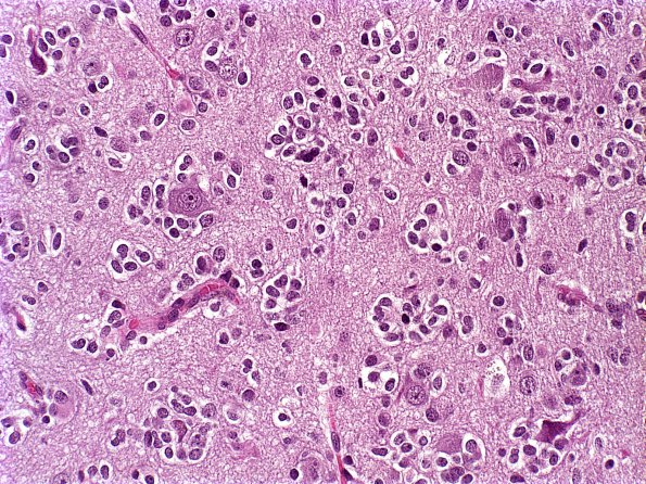
Washington University Experience | NEOPLASMS (GLIAL) | Oligodendroglioma - IDH1 mutant, del 1p19q - Grade 3 | 15A Oligodendroglioma, anaplastic (Case 15) b
Case 15 Histories ---- The patient is a 61 year old man with a right frontal lobe mass that was first biopsied in 1996. It recurred 4 years later. His disease is currently stable, with no radiologic evidence for recurrence. ---- 15A The tumor cells are composed predominantly of round uniform nuclei with bland chromatin and clear perinuclear halos. A rich branching capillary network is seen in the background. There is prominent perineuronal satellitosis seen in H&E. Microcystic spaces and hypercellular nodules are seen in some areas. The mitotic index is brisk. Although there is no endothelial hyperplasia in the 1996 specimen, it is prominent in the 2000 specimen. Likewise, no necrosis is seen in the 1996 specimen, though there are large zones of coagulative necrosis in the 2000 recurrence. This is associated with fibrin deposition and hyalinized vessels, suggestive of radiation necrosis. ---- Not shown: The tumor cells are negative for Neu-N. They are also negative for synaptophysin in the 1996 specimen, though patchy immunoreactivity is seen in the 2000 specimen. The morphologic and immunohistochemical features of both specimens are consistent with anaplastic oligodendroglioma, WHO grade 3. The focal immunoreactivity for synaptophysin in the recurrent specimen does not preclude this diagnosis since it has been described in a subset of oligodendrogliomas. ---- In the 1996 specimen, deletions of both 1p and 19q were identified. Similarly, the 2000 specimen showed evidence for relative deletions of both 1p and 19q.

