Table of Contents
Washington University Experience | NEOPLASMS (GLIAL) | Oligodendroglioma - IDH1 mutant, del 1p19q - Grade 3 | 18A Oligodendroglioma, anaplastic, garlands (WHO III, Case 18) H&E 14 copy.jpg
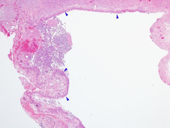
18A The featured histopathology in this case is the proliferation of subpial aggregation of tumor cells as well as prominent microvascular proliferation in garland-type structures. A mitotic count performed on slide C2 yields 13 mitotic figures per 10 high power fields. Calcifications are scattered throughout. ---- Not shown: An immunostain for the IDH-1 mutant protein (p. R132-H) is positive. An Olig-2 stain is strongly and diffusely positive in tumor cells. An immunostain for synaptophysin is positive in tumor cells. The expression of synaptophysin suggests an entity which has been previously described as "oligodendrogliomas with neurocytic differentiation" (PMID: 12430711), in which four cases of both low and high-grade oligodendrogliomas showed areas of neurocytic differentiation; all showed at least partial immunoreactivity for synaptophysin, and three of four cases showed codeletion of chromosomal arms 1p and 19q. The prognostic significance of neuronal/neurocytic differentiation in oligodendrogliomas, if any, is unclear. Overall, given the elevated mitotic index, microvascular proliferation, and the presence of significant nuclear atypia, this tumor is best classified as an anaplastic oligodendroglioma, WHO 3, and likely represents progression from the patient's previously diagnosed oligodendroglioma, WHO grade 2, in the same location.

