Table of Contents
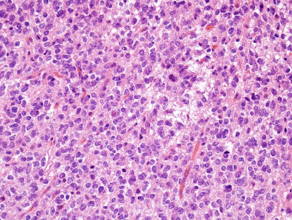
Washington University Experience | NEOPLASMS (GLIAL) | Oligodendroglioma - IDH1 mutant, del 1p19q - Grade 3 | 4A1 Oligodendroglioma, anaplastic WHO Grade 3, Case 4) H&E 2
Case 4 History ---- The patient is a 54 year old woman with a history of seizures. Per clinical records, the patient underwent excision of a right frontal cerebral lesion at an OSH in 1998 at the age of 41. She subsequently underwent excision of a recurrent lesion in 2004.---- 4A1-3 The neoplasm forms sheets of regularly spaced cells. The neoplastic cells have ovoid to round nuclei with highly irregular nuclear contours, notching, and variable chromatin ranging from hyperchromatic to stippled. The background vasculature shows lumens with irregular walls and dilatation; however, endothelial cells remain single-layered, i.e., frank endothelial hyperplasia is not present. Necrosis is not observed. Portions of the specimen are markedly hypercellular and the mitotic index in these areas numbers 7 mitoses/10HPF.

