Table of Contents
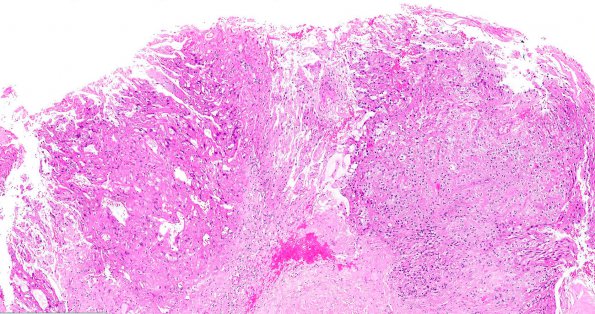
Washington University Experience | NEOPLASMS (GLIAL) | PLNTY | 2D1 PLNTY (Case 2, Resection 2) H&E 5X
Case 2, Resection #2 (2011) ---- 2D1-2 Multiple small fragments of neoplastic tissue are markedly edematous, featuring tumor cells with small-to-medium atypical hyperchromatic nuclei, moderate amounts of eosinophilic cytoplasm, and irregular wispy cell processes that form a sparse meshwork with fine, inconspicuous capillaries and a few larger vessels; small subtle collections of tumor cell processes surround some vessels, consistent with pseudorosette formation. One piece of more solid tissue exhibits a few different histological patterns. In one focus, the tumor cells appear ovoid with smaller oval atypical nuclei, and cleared or microvacuolated cytoplasm; in a nearby focus, similar tumor cells feature modest bellies of eosinophilic cytoplasm. A few vessels exhibit mural thickening and hyalinization. Only two adjacent mitotic figures are identified in a significant amount of tissue. There is no definitive evidence of microvascular proliferation, endothelial hyperplasia, or necrosis. (H&E)

