Table of Contents
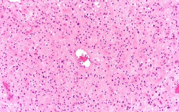
Washington University Experience | NEOPLASMS (GLIAL) | PLNTY | 2F1 PLNTY (Case 2, Resection 3) H&E 20X 1
Case 2, Resection #3 (2019) ---- In February 2019, the patient was admitted after a witnessed generalized tonic-clonic seizure, which led to a subarachnoid hemorrhage. Follow-up brain MRI on July 2019 showed progressive interval increase in the non-enhancing diffusion restricted heterogeneous mass at the left ventricle trigone. Operative procedure: Left parietal twist drill burr hole for frameless stereotactic needle biopsy and laser interstitial thermal therapy (LITT) of recurrent glioneuronal tumor. ---- 2F1-4 The 2019 neoplasm appeared low-grade with a predominantly glial phenotype harboring a solid growth pattern. The tumor cells demonstrate relatively bland-appearing round nuclei with generally smooth chromatin and crisp nuclear contours. There is some degenerative type nuclear atypia as well. The cells are embedded in a fibrillary background with generally indistinct cell borders harboring a perivascular pseudo-rosetted architecture focally. Small clusters of tumor cells also possess clear cytoplasm with an 'oligodendroglial' appearance. The tumor vasculature is mostly composed of thin-walled capillaries with no evidence of microvascular proliferation. Mitoses are hard to find and necrosis is not appreciated either. Rosenthal fibers, eosinophilic granular bodies, and/or microcalcifications are also not apparent.

