Table of Contents
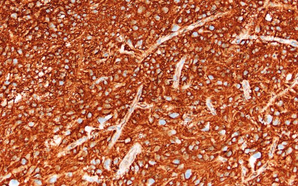
Washington University Experience | NEOPLASMS (GLIAL) | PLNTY | 2I3 PLNTY (Case 2, Resection 3) CD34 20X 3
Tumor cells are intensely immunoreactive for CD34. ---- Ancillary data (not shown): There was occasional perinuclear dot-like positivity for synaptophysin. EMA is negative through-out. Neurofilament highlights only rare axons. Mutant BRAF (V600E) is negative. Ki-67 (MIB-1 antibody) highlights a very low proliferation index of less than 1%. ---- FISH showed no evidence of EGFR amplification and/or PTEN loss. Additional FISH testing for BRAF (7q34) Break-Apart (performed at Cincinnati Children's Hospital) is “abnormal and consistent with three copies of BRAF (7q34) fusion”. ---- Comment: The collective morphological, immunohistochemical and cytogenetic findings are consistent with an integrated diagnosis of "low-grade glioneuronal neoplasm with predominantly glial component, BRAF altered". Specifically, the 'neuronal' differentiation in the current biopsy is rather limited. Since the tumor is morphologically not fitting into a distinct diagnostic category, we preferred to use the term "low-grade glioneuronal neoplasm with predominantly a glial component" and did not assign a specific WHO grade. Of note, BRAF fusion, as detected in the current case, is not pathognomic of a specific diagnosis but is generally associated with a significant number of low-grade CNS tumors particularly in pediatric setting, that predominately correspond to WHO grade I designation. The very low proliferation indices as well as solid nature of the current tumor appear to fit with that notion. However, we also make a note of the ventricular involvement by this tumor and clinical history of local recurrences.

