Table of Contents
Washington University Experience | NEOPLASMS (GLIAL) | PLNTY | 3A1 PLNTY (Case 3, Resection 1) FLAIR - Copy copy
Case 3, History (2 resections) ---- The patient was a 6-year-old girl who presented with new onset seizures. MRI studies in March 2018 showed a 0.8 x 1.5 x 0.7 cm non-enhancing, FLAIR-signal suppressing, cystic lesion with internal septation within the left middle temporal gyrus, without diffusion restriction or susceptibility artifact. Radiological impression: Main considerations include Dysembryoplastic neuroectodermal tumor (DNET), pleomorphic xanthoastrocytoma (PXA), and ganglioglioma. Operative procedure: Robotic stereotactic assisted (ROSA) left temporal lesion biopsy with subsequent laser induced thermal therapy (LITT). The biopsy confirmed the presence of a glioneuronal neoplasm. ---- At the time of her second resection in 2020, the now 8 year old patient was improved with her spells lasting 15-20 seconds, however they remained refractory to medication. A stereo EEG implantation confirmed seizure onset in the perilesional region of the inferior posterior temporal lobe. Operative procedure in 2020: Left sided temporal craniotomy for resection of seizure focus with frameless stereotactic neuro navigation microsurgical dissection. ----
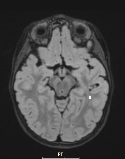
Case 3, Resection #1 (2018) ---- A1-4 MRI studies: ---- 3A1 A hypointense lesion (arrow) is identified in the left temporal lobe in this FLAIR study.

