Table of Contents
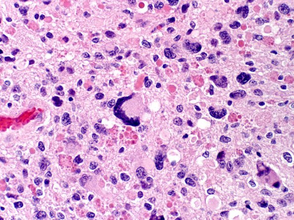
Washington University Experience | NEOPLASMS (GLIAL) | Pleomorphic Xanthoastrocytoma (PXA) | 10A1 PXA & GG (Case 10) H&E 3
Case 10 History ---- The patient is an 18 year old girl who presented with seizures. Imaging revealed a superficially located right parietal cortical mass. ---- 10A1-4 Sections reveal a moderately cellular glial neoplasm centered in the cortex. The majority of the tumor cells have a spindled morphology arranged in fascicles and a storiform pattern. Astrocytic-appearing cells are also encountered with variable quantities of eosinophilic cytoplasm. The tumor nuclei range from spindled to irregular and hyperchromatic, with scattered multinucleated giant cells. There are numerous eosinophilic granular bodies and occasional Rosenthal fibers. Perivascular and intratumoral lymphocytic accumulation is identified. At the edge of the tumor, there is a degree of cortical infiltration. Microcalcifications are seen focally.

