Table of Contents
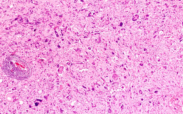
Washington University Experience | NEOPLASMS (GLIAL) | Pleomorphic Xanthoastrocytoma (PXA) | 11A1 PXA (Case 11) 10X
Case 11 History ---- The patient is a 13 year old boy who has a cystic, partially calcified, heterogeneous right temporal lobe mass with minimal enhancement, measuring 3.5 cm in diameter, and a right parietal superficial mass, measuring 1.5 cm in diameter with mild enhancement and erosion of the inner table of the skull. ---- 11A1-6 This is an astrocytic neoplasm with a fascicular architecture. Pleomorphic, multinucleated giant cells with bizarre cytologic features are seen, along with occasional large xanthomatous cells showing intracellular accumulation of lipid. Eosinophilic granular bodies and perivascular lymphocytes are also present. Necrosis is absent, and mitoses are difficult to identify.

