Table of Contents
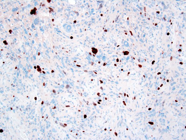
Washington University Experience | NEOPLASMS (GLIAL) | Pleomorphic Xanthoastrocytoma (PXA) | 11E PXA (Case 11) Ki67 1.jpg
Ancillary data (not shown): Immunohistochemical staining for synaptophysin shows rare, positive cells. NeuN is negative within tumor cells. Neurofilament immunoreactivity highlights the focally infiltrative nature of the tumor. The proliferative MIB-1 index in this case is somewhat higher than that typically seen with a WHO Grade 2 PXA; nonetheless, the more reliable low mitotic count has caused us to make a diagnosis of pleomorphic xanthoastrocytoma with increased proliferative index WHO Grade 2.

