Table of Contents
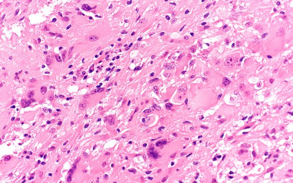
Washington University Experience | NEOPLASMS (GLIAL) | Pleomorphic Xanthoastrocytoma (PXA) | 12A1 PXA (Case 12) H&E 40X 1
Case 12 History ---- The patient is a 51 year old man with a history of neurofibromatosis type I who presented with headaches. Imaging showed a 4.0 x 4.1 x 3.9 cm mixed solid and cystic enhancing lobulated mass within the right frontal lobe and right corpus callosum. Operative procedure: Stealth guided brain biopsy. ---- 12A1,2 This is a hypercellular neoplasm consisting of neoplastic cells with a varied morphological appearance with a sheet-like growth pattern. Many of the neoplastic cells have abundant opalescent eosinophilic cytoplasm and enlarged, irregularly-shaped markedly pleomorphic hyperchromatic nuclei. These cells form a 'pavemented' pattern with respect to one another and have highly irregular cell-cell borders. Occasional cells also have vacuolated cytoplasm, i.e., are xanthomatous. Multinucleated giant cells are also present. Cells with elongated hyperchromatic nuclei and fibrillary cytoplasmic processes are also present. Eosinophilic granular bodies are identified focally. Mitotic figures appear infrequent, numbering up to 3/10 HPF fields.

