Table of Contents
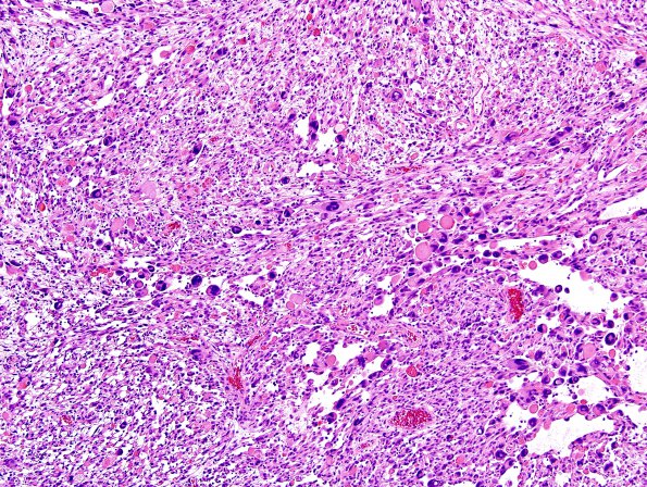
Washington University Experience | NEOPLASMS (GLIAL) | Pleomorphic Xanthoastrocytoma (PXA) | 15A1 PXA (Case 15) H&E 9
Case 15 History ---- The patient is a 29 year old man with an intra-axial partially cystic and enhancing mass (4.1 x 3.5 x 2 cm) in the left temporal lobe. Operative procedure: resection. ----15A1-4 This is a relatively solid-appearing glial neoplasm with sharp demarcation from adjacent brain, but focal perivascular spread for a short distance beyond the main tumor mass. Perivascular lymphocytic cuffing is prominent in some areas. There is moderate to focally marked nuclear pleomorphism, with most of the tumor cells containing elongate mildly hyperchromatic nuclei. However, bizarre multinucleated forms are also evident. The majority of tumor cells contain abundant eosinophilic cytoplasm and occasional cells contain clear vacuolated cytoplasm, consistent with lipid accumulation (xanthoastrocytes). There are numerous eosinophilic granular bodies throughout the tumor. Mitotic figures are hard to find and there is no definite evidence of microvascular proliferation or necrosis.

