Table of Contents
Washington University Experience | NEOPLASMS (GLIAL) | Pleomorphic Xanthoastrocytoma (PXA) | 16A1 PXA WHO II (Case 16) H&E 13.jpg
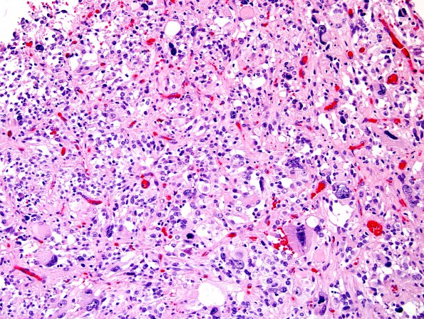
Case 16 History ---- This is a 22 year old man with a left temporal mass. Preoperative MRI showed a large solid and cystic expansile mass centered in the anterior left temporal lobe with associated mass effect and mild left to right midline shift with central irregular foci of enhancement and elevated relative cerebral blood volume. ---- 16A1-9 Microscopic examination revealed a hypercellular neoplasm composed of medium to large cells with abundant eosinophilic cytoplasm and focal prominent nuclear pleomorphism. Tumor cells with an epithelioid appearance and cytoplasmic filamentous material are readily seen. Several multinucleated tumor cells and cells with lipidized cytoplasm are present. The tumor shows a significant leptomeningeal component with infiltration between superficial blood vessels that show marked hyalinization. No significant mitotic activity is seen. Necrosis or vascular proliferation are not seen. The surrounding cortex shows gliosis with focal infiltration by tumor cells. Perivascular and intratumoral aggregates of lymphocytes are seen. Rare eosinophilic granular bodies are present and highlighted by PAS stain.

