Table of Contents
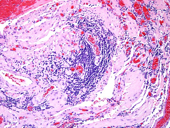
Washington University Experience | NEOPLASMS (GLIAL) | Pleomorphic Xanthoastrocytoma (PXA) | 16A2 PXA WHO II (Case 16) H&E 10.jpg
Microscopic examination revealed a hypercellular neoplasm composed of medium to large cells with abundant eosinophilic cytoplasm and focal prominent nuclear pleomorphism. Tumor cells with an epithelioid appearance and cytoplasmic filamentous material are readily seen. Several multinucleated tumor cells and cells with lipidized cytoplasm are present. Lymphocytic infiltration is common. (H&E)

