Table of Contents
Washington University Experience | NEOPLASMS (GLIAL) | Pleomorphic Xanthoastrocytoma (PXA) | 16A9 PXA WHO II (Case 16) H&E 2.jpg
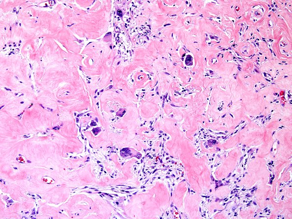
Collagenized vessels are focally encountered. (H&E) ---- Ancillary data (not shown) Pericellular reticulin deposition is focally appreciated. Lipid droplets were documented within tumor cells by Oil Red O stain. The tumor cells are strongly and diffusely positive for GFAP immunostain. lmmunohistochemical stain for synaptophysin highlights cortical neuropil and rare tumor cells. Neurofilament immunohistochemical stain is negative in most of the tumor and highlights peripheral entrapment of axons. CD34 immunostain highlights blood vessels but not reticulated forms. EMA is negative. Ki-67 proliferation index is up to 5% (significant non-tumor positive nuclei). An immunohistochemical stain for p53 is positive in approximately 6% of tumor nuclei. The tumor is negative for BRAF V600E mutation ---- A diagnosis of PXA Grade 2 was made.

